- HomeElectronicsCamera & PhotoBinoculars & ScopesMicroscopesCompound MicroscopesCompound Trinocular Microscopes
The AmScope T590A-DK compound trinocular microscope has a Siedentopf binocular mount with interchangeable pairs of 10x widefield and 16x widefield eyepieces, a reverse-mounted nosepiece with four DIN achromatic high-contrast objectives, Brightfield/Darkfield halogen illumination, a double-layer mechanical stage with a stage stop to protect slides and objectives from damage, and anti-mold surfaces to preserve optics in high-humidity areas. The trinocular head has a vertical camera mount and a Siedentopf binocular mount with interchangeable pairs of WF10x and WF16x eyepieces with 55 to 75mm Siedentopf interpupillary adjustment, a fixed 30-degree vertical inclination to reduce eye and neck strain, and a 360-degree rotation capability to provide a more comprehensive view and enable sharing. Dioptric adjustment accommodates individual eye-strength differences. The vertical trinocular port accepts a camera with a 23mm or C-Mount adapter (camera sold separately). The reverse-mounted revolving nosepiece has 4x, 10x, and 40xS, and 100xS (oil) DIN achromatic high-contrast objectives that provide color correction of magnified images. High-contrast objectives provide sharp color distinction. The 40xS objective is spring loaded to prevent slide damage when focusing. The 100xS spring-loaded oil-immersion objective uses oil between the specimen and the objective lens to provide increased resolution over a standard objective. A compound microscope is used for inspection and dissection of specimens when two-dimensional images are desired.
The microscope has lower (transmitted, diascopic) Brightfield/Darkfield (BF/DF) illumination that transmits light up through the specimen for enhanced visibility of translucent and transparent objects. Brightfield (BF) illumination allows the specimen to absorb light, resulting in a dark image on a light background, and Darkfield (DF) illumination reflects light off the specimen, resulting in a light image on a dark back
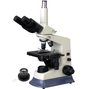
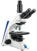
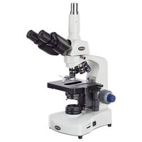
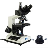
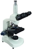
AmScope T590A-DK Professional Compound Trinocular Microscope, WF10x and WF16x Eyepieces, 40X-1600X Magnification, High-Contrast Objectives, Brightfield/Darkfield, Halogen Illumination, Abbe Condenser, Double-Layer Mechanical Stage, Anti-Mold, 110V
The AmScope T590A-DK compound trinocular microscope has a Siedentopf binocular mount with interchangeable pairs of 10x widefield and 16x widefield eyepieces, a reverse-mounted nosepiece with four DIN achromatic high-contrast objectives, Brightfield/Darkfield halogen illumination, a double-layer mechanical stage with a stage stop to protect slides and objectives from damage, and anti-mold surfaces to preserve optics in high-humidity areas. The trinocular head has a vertical camera mount and a Siedentopf binocular mount with interchangeable pairs of WF10x and WF16x eyepieces with 55 to 75mm Siedentopf interpupillary adjustment, a fixed 30-degree vertical inclination to reduce eye and neck strain, and a 360-degree rotation capability to provide a more comprehensive view and enable sharing. Dioptric adjustment accommodates individual eye-strength differences. The vertical trinocular port accepts a camera with a 23mm or C-Mount adapter (camera sold separately). The reverse-mounted revolving nosepiece has 4x, 10x, and 40xS, and 100xS (oil) DIN achromatic high-contrast objectives that provide color correction of magnified images. High-contrast objectives provide sharp color distinction. The 40xS objective is spring loaded to prevent slide damage when focusing. The 100xS spring-loaded oil-immersion objective uses oil between the specimen and the objective lens to provide increased resolution over a standard objective. A compound microscope is used for inspection and dissection of specimens when two-dimensional images are desired.
The microscope has lower (transmitted, diascopic) Brightfield/Darkfield (BF/DF) illumination that transmits light up through the specimen for enhanced visibility of translucent and transparent objects. Brightfield (BF) illumination allows the specimen to absorb light, resulting in a dark image on a light background, and Darkfield (DF) illumination reflects light off the specimen, resulting in a light image on a dark back
in next 2 weeks
By clicking "Accept All", you consent to our use of cookies.